News
LSU recently upgraded its Scanning Electron Microscope (SEM) with new AZtec software, making it easier to see and analyze materials at the nanoscale. The upgrade allows researchers to scan large areas, like entire rock thin sections, while still capturing fine details. Led by geology professors Eirini Poulaki and Brandon Shuck, this improvement helps scientists quickly identify important minerals and create high-resolution chemical maps. The new system also supports 3D analysis using machine learning and X-ray data, giving students and researchers more powerful tools for geoscience and materials research.
You can learn more about this upgrade on the LSU Science Next blog.


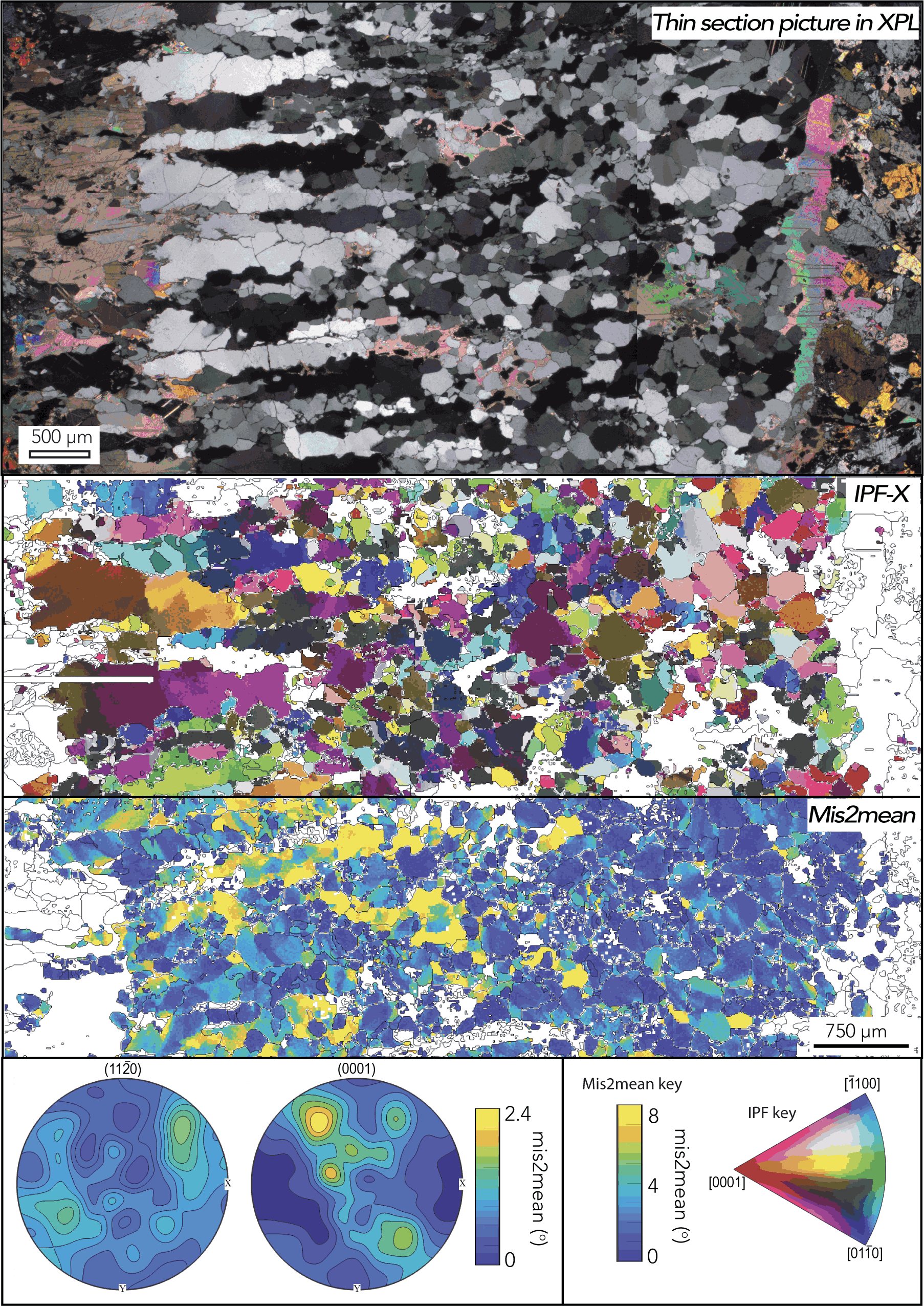
Top panel: Thin section picture of quartz vein in cross polarized light. Mis2mean: Map of misorientations mapped by the large area Aztec software capabilities that LSU now has access to. This map shows the internal misorientation within the quartz grain, showing how much each point deviates from the grains' average orientation. IPF-X: A map that shows the crystallographic orientation of grains; different colors represent different quartz crystal orientations. Bottom panel: Pole figures showing crystal prefer orientation along the c and a quartz axis and legend.
All EBSD data shown in this figure were collected at the Molecular Analysis Facility at the University of Washington using equipment similar to that at LSU, which now has the capability to obtain the same datasets.
Congrats to Daniel Geldof!
Daniel’s master’s research in Dr. Prosanta Chakrabarty’s lab on the rockhead poacher just made a big splash! His study was featured in Science and even in The New York Times. Daniel explored the idea that this quirky fish may use its unusual head cavity to
produce sound.
To dive into one of nature’s strangest fish skulls, he used our cutting-edge Thermo Fisher HeliScan microCT Mark II, showing how our core facility supports exciting, high-impact research.


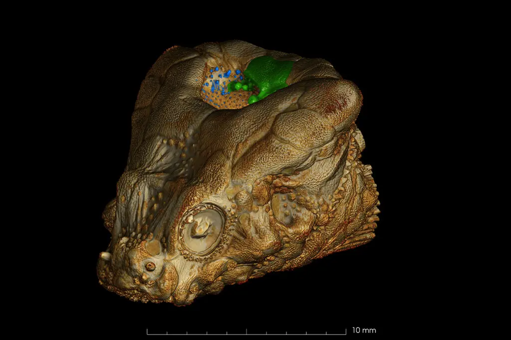
Micro-CT model of the rockhead poacher (Bothragonus swanii). Scanned with AMAC’s ThermoFisher HeliScan MKII. Fish stained with phosphomolybdic acid.
Big news from LSU!
The Advanced Microscopy and Analytical Core has installed the Thermo Fisher Spectra 300 STEM, the most advanced microscope in Louisiana, made possible through a $10 million investment from the U.S. Army Research Office.
This state-of-the-art instrument enables researchers to visualize materials at the atomic scale, driving breakthroughs in materials science, biomedicine, and national defense. With this powerful technology, LSU is leading Louisiana into a new era of innovation.
Learn more about this major milestone on Louisiana First News and the LSU Blog.
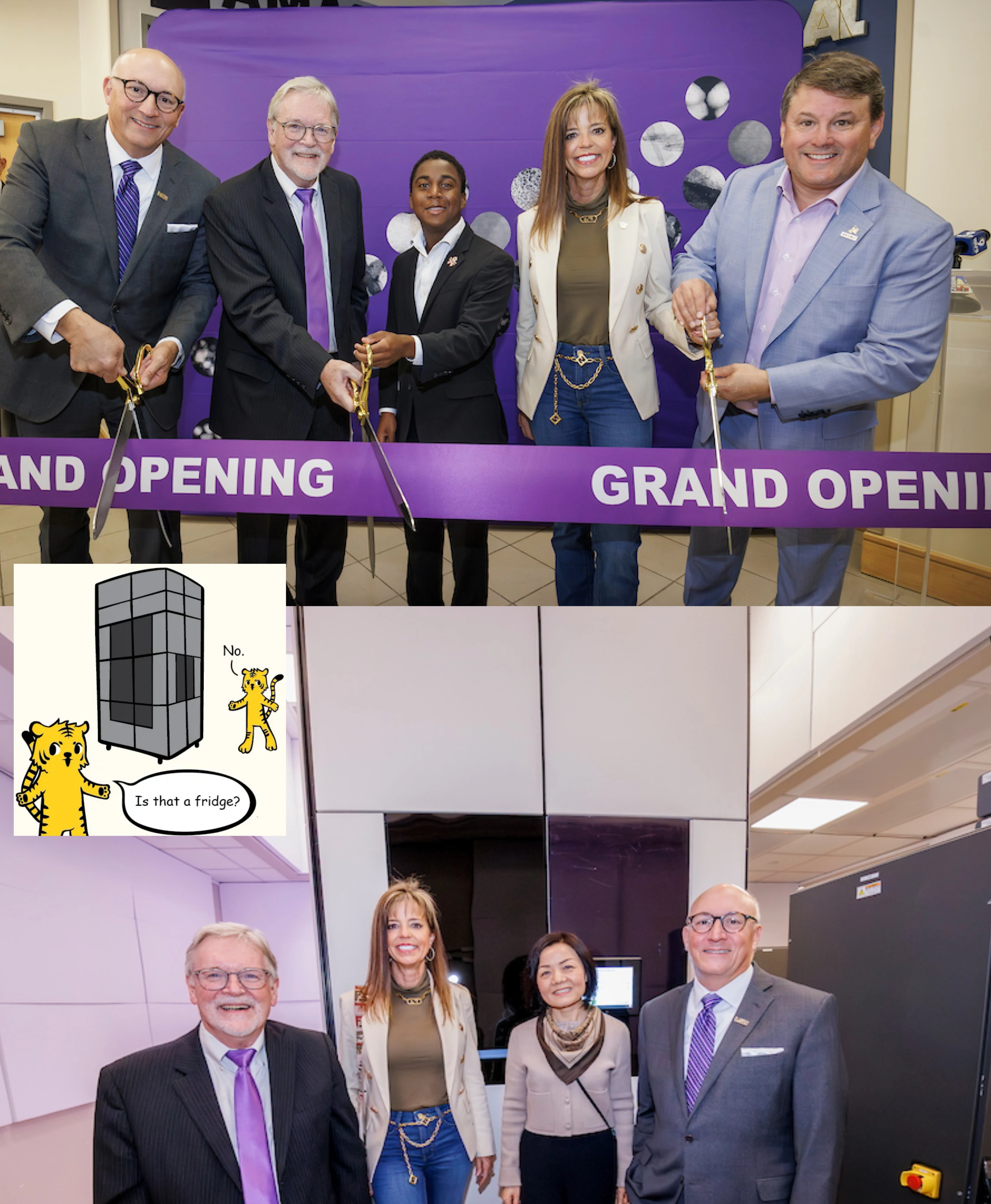
Photos by Eddy Perez, art by Brittany Ball and Elsa Hahne.
Congratulations to Amaya Wanniarachchi!
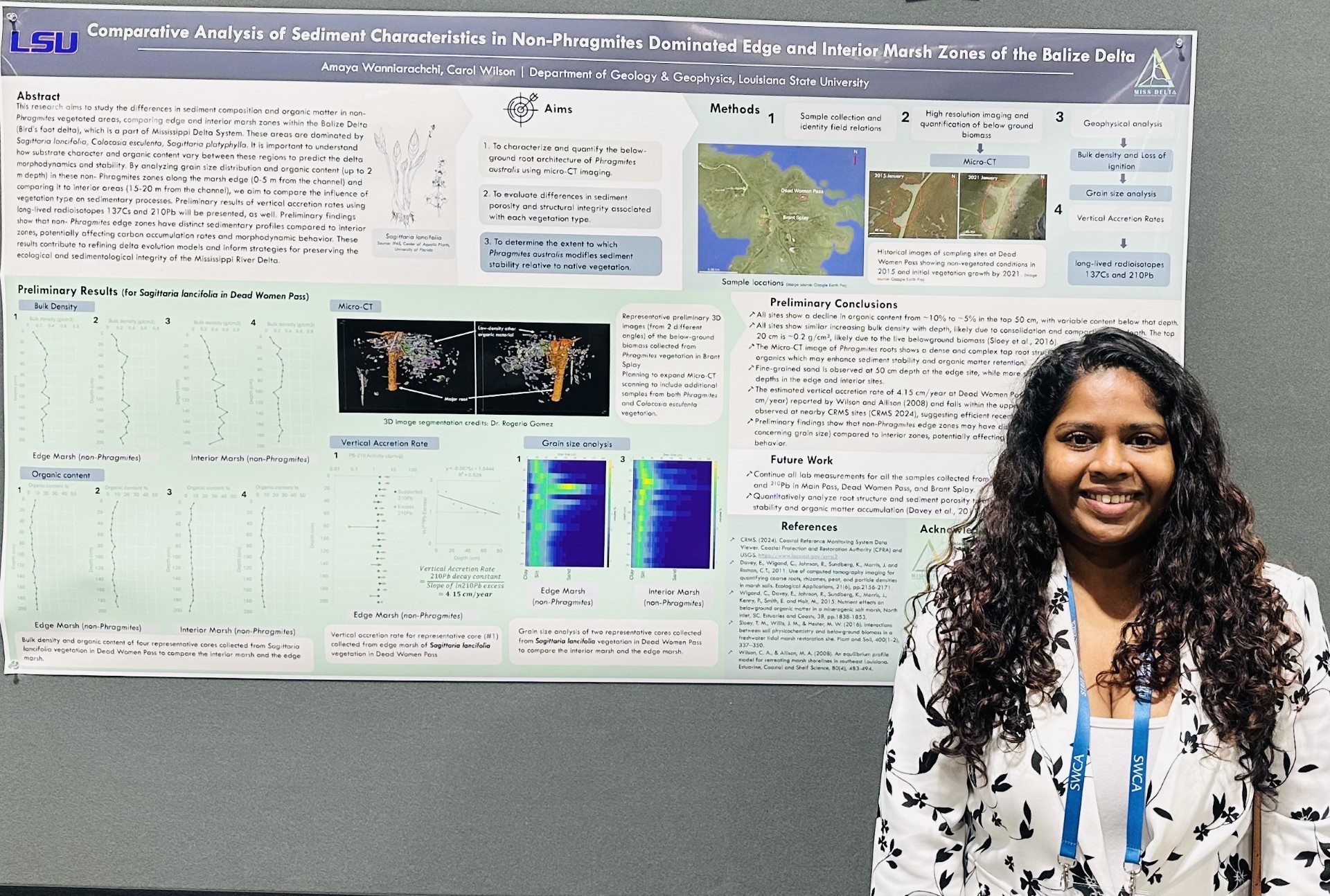
Amaya, a PhD student in Dr. Carol Wilson’s lab, won third place in the graduate student category of the poster competition at State of the Coast 2025.
Her award-winning poster featured several exciting results, including a 3D image captured using our Thermo Fisher HeliScan microCT Mark II.
We’re proud to be supporting this outstanding research!


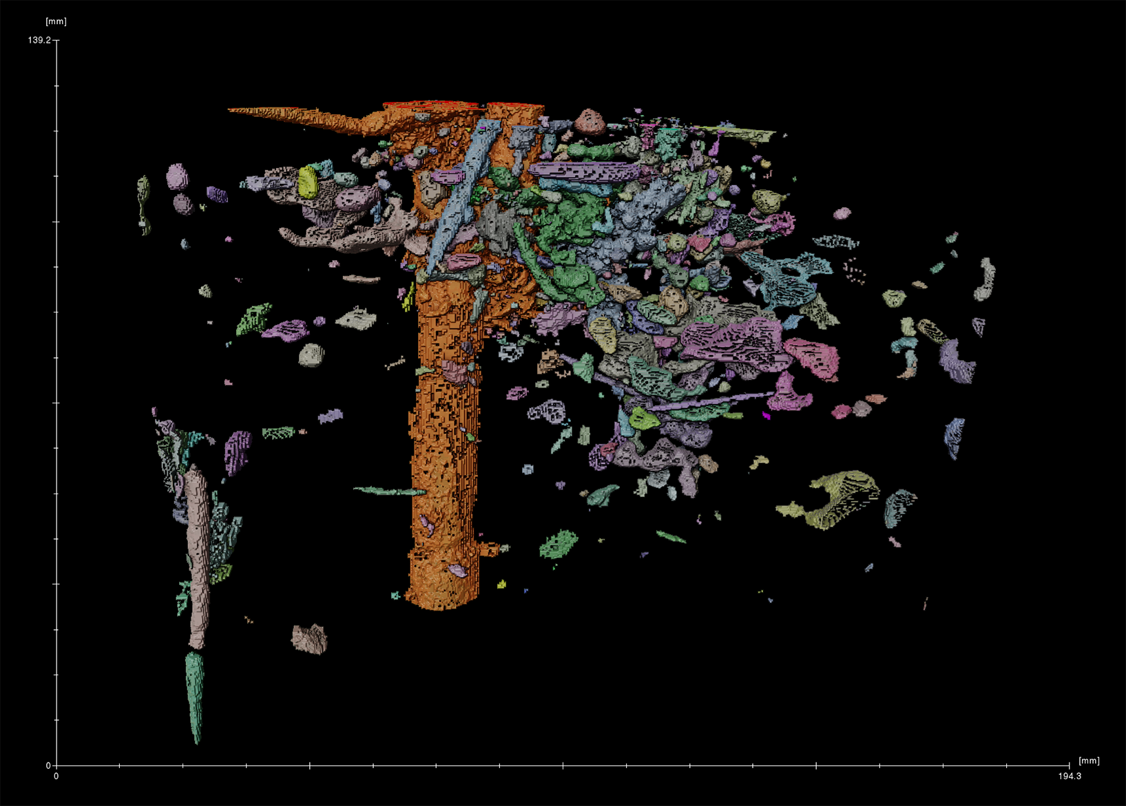
Preliminary 3D reconstruction from microCT imaging of below-ground biomass sampled from Phragmites stands in the Brant Splay region.
Congrats to Vishakha Vishwakarma!
Vishakha, a former PhD student from Dr. SeYeon Chung’s lab, was one of the winners of the 2023 Huygens Image Contest!
Her stunning image, captured on our Leica TCS SP8 Confocal Microscope with a White Light Laser (WLL), earned her the 6th place spot. We're proud to have been part of this amazing work!

 .
.
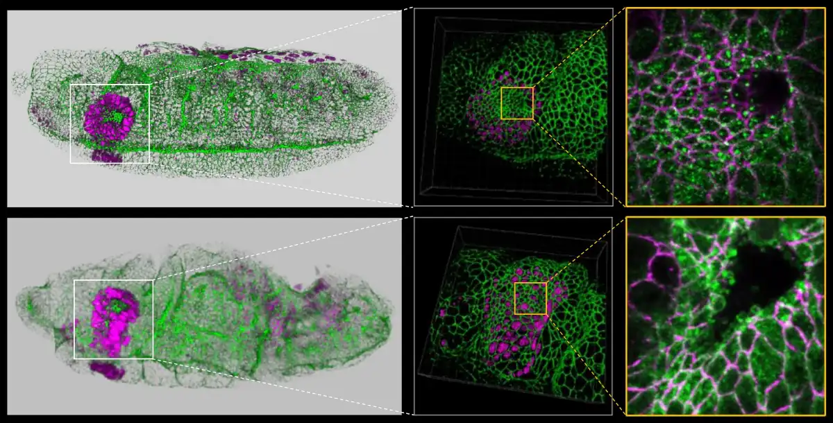
Clear visualization of interesting phenotype. Unlike wild-type embryo (top panels), loss of Smog GPCR (bottom panels) results in slight disruption of the overall embryonic morphology, including defects in the head region, elongated salivary gland placode, a wavy embryo surface and an irregular ventral midline. 3D reconstructed images (middle panels) show the salivary gland placode elongated along the DV axis and enlarged cells upon loss of smog. Strikingly, smog loss also results in the formation of numerous blebs in the apical membrane of salivary gland cells, which are not observed in wild-type salivary gland cells, suggesting defects in the cortical actin network (rightmost panels). Overall, our work suggests multifunctional roles of GPCR signaling during epithelial morphogenesis.
An image acquired using the Leica TCS SP8 Confocal Microscope with White Light Laser (WLL) at our microscopy facility was featured on the cover of Biology Open, as part of the publication 'Nucleolar stress in Drosophila neuroblasts, a model for human ribosomopathies' by Sonu S. Baral, a former doctoral student in Dr. Patrick J. DiMario’s laboratory.


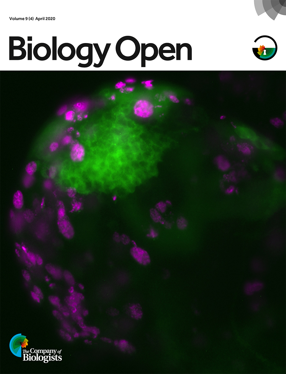
In a study by Baral and colleagues wild-type (w1118) larval brains at 3 days old were EdU pulse labelled (Click-iT Alexa Fluor 594) for 30 min to visualize all S-phase cells: neuroblasts (NB) and ganglionic mother cells (GMCs). This merged confocal image shows EdU-labelled cells (magenta; mushroom body NBs and their GMCs) nestled within the GFP-labelled mushroom body NBs lineage (green) near the posterior regions of the central brain lobe. Scale bar: 50 microns. Image licensed under a Creative Commons Attribution 4.0 International license.